AI in Images: From Ghibli Animations to Your Lung X-rays
Explore how generative AI evolved from artistic creations like Ghibli-style images to transforming medical imaging for accurate, fast diagnosis in healthcare.

The Evolution from Generative Text to Precision Medical Imaging
Over the past few years, generative AI has worked its way from producing simple text to creating and reading complex images. Many people know about the copyright issues between OpenAI and the Ghibli Studio which caused AI image-generation tools to be unavailable for a little while. Regardless of the challenges listed above, generative AI technology has made steady progress and now produces and interprets medical images accurately.
Medical experts and researchers have recognized that AI in imaging has a significant impact. Many hospitals and laboratories use AI to support their clinical work such as by improving diagnostics, simplifying processes and reducing errors from human interpretation.
Clinical Image Interpretation: Enhancing Accuracy and Efficiency through AI
AI is used in various areas of medicine, leading to better results in diagnosing and treating patients. Thanks to AI, chest radiography which is commonly performed, can quickly find pneumonia (such as a tool called MedCheX, which is developed by a Taiwanese team), tuberculosis, tumors and other abnormalities. By using these AI systems, a patient’s screening and diagnosis can speed up so that results are used sooner.
In both obstetrics and cardiology, AI-assisted ultrasound scans are accurate in measuring body parts, noticing any birth defects and evaluating the heart’s functions in the womb. Besides, scans done with computed tomography scan (CT) and Positron Emission Tomography (PET) produce a lot of detailed data. Using AI, deep learning algorithms can swiftly and accurately review large amounts of image slices to spot tiny lesions missed by humans.
To assess a patient’s tumor status and understand the disease, immunohistochemical (IHC) techniques are used heavily in pathology laboratories. Typically, evaluating samples under the microscope with personal judgments introduced variability and could lead to false-positives, so many tests used double-blind methods to reduce errors. Thanks to AI, cells can now be examined and interpreted by the computer, making it possible to improve accuracy and minimize any inconsistencies.
Image Analysis: From ImageJ Macros to AI Automation
In the past, scientists used ImageJ and similar platforms to ease the work of analyzing microscopy images automatically with macros. While it proved to be a good solution, it placed major obstacles on the path of researchers without programming skills. Looking through past research and GitHub required a lot of time, with macros often being either unsafe, impossible to combine with other software or simply not suitable for researchers’ needs.
With the help of AI-driven systems, people with limited coding background can easily start using analysis pipelines for their research. Thanks to intuitive tools and AI, scientists are able to speed up the measurement of fluorescence images and look at cell shape. From the results I have seen, using an AI technique for analysis led to far better outcomes and more dependable results. This made our research progress much faster and more trustworthy.
Pharmaceutical Giants Leveraging AI: Roche’s Breakthrough VENTANA TROP2 Device
To benefit from new techniques, Roche and other pharmaceutical companies now use AI in inventing better tools for diagnosis. On April 29, 2025, Roche introduced a new tool called VENTANA TROP2 (EPR20043) RxDx Device to provide treatment-guiding information for people diagnosed with advanced non-small cell lung cancer (NSCLC).
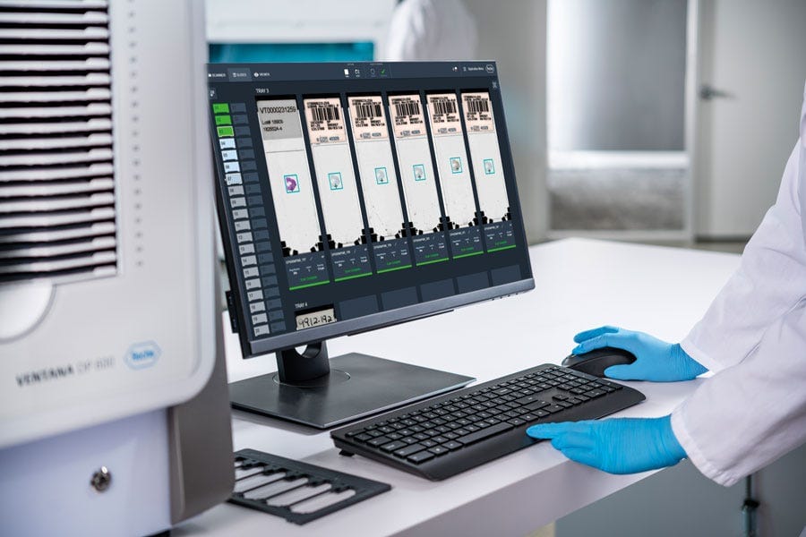
VENTANA TROP2 is meaningful because it is the first AI-assisted technology of its sort to receive the “Breakthrough Device Designation” from the FDA. As a result of this designation, patients can get the device quicker as the approval process is only slightly delayed. The system has Roche’s modern staining system (BenchMark ULTRA), specific detection techniques (OptiView DAB), top-quality scanners and software called the navify Digital Pathology Image Management System.
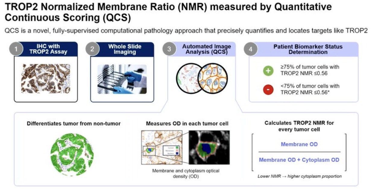
Initially, scientists stain a sample of the cancer tissue to look for a protein called TROP2. After staining, the AI system created by AstraZeneca looks at the digital images of the tissue samples it has provided. The software assesses the degree of staining to provide a ranking as the Normalized Membrane Ratio or NMR. The pathologist tests the staining and identifies the immunohistochemistry-stained cells matching the tumor before the AI system classifies the patient outcome as either TROP2-positive or negative.
Since AI automates this evaluation, it eliminates the need to evaluate tissue samples by sight and ensures that results are dependable, precise and always the same. As a result, doctors can accurately determine which patients are suitable for a drug such as datopotamab deruxtecan which is made for treating cancers that show the TROP2 marker. For these patients, it becomes especially important since they do not have the usual genetic changes identified in tumor treatment.
Looking Forward: The Future of AI in Medical Imaging and Precision Healthcare
AI is making a big impact in medical imaging, as well as in entertainment and creative industries. AI-powered tools are helping healthcare experts to diagnose patients quickly, provide effective monitoring services and use targeted treatment plans.
When disciplines work together, AI will have even more influence as its algorithms develop. Soon, AI-supported tools will be crucial for making decisions in healthcare, developing new medicines and implementing precision medicine. If health services embrace the capabilities of AI, they will be able to discover new medical solutions and improve the wellbeing of patients worldwide.


